Original Article
HJOG 2026, 25 (4), 300-314 | doi: 10.33574/hjog.0638
Tsorva Erikaiti1, Kakourou Georgia2, Christopikou Dimitra3,5, Davies Stephen1, Zacharia Stavroula1, Sfontouris Ioannis1,4, Makrakis Evangelos1,4, Frysira Helen2, Tzetis Maria2, Mastrominas Minas1, Traeger-Synodinos Joanne2
1Hygeia IVF Embryogenesis Unit, Athens, Greece
2Laboratory of Medical Genetics, St. Sophia’s Children’s Hospital, Medical School, National and Kapodistrian University of Athens, Greece
3Assisting Nature IVF Unit, Thessaloniki, Greece
4Department of Basic and Clinical Sciences, Medical School, University of Nicosia, UNIC Athens, Greece
5IASO Institute of Life IVF Unit, Athens, Greece
Correspondence: Tsorva Erikaiti, PGT Lab Director , Hygeia IVF Embryogenesis, Athens, Greece, email: etsorva@hygeiaivf.gr

Abstract
Background: Embryo selection remains one of the principal determinants of success in assisted reproductive technology (ART). Current selection methods rely primarily on morphological assessment, which provides only indirect and imperfect prediction of embryo developmental competence and implantation potential. Identification of reliable non-invasive molecular biomarkers reflecting oocyte competence represents a major objective in reproductive medicine.
Apoptosis is a programmed cell death process essential for tissue homeostasis and is closely associated with key reproductive events, including follicular atresia. MicroRNAs (miRNAs) regulate gene expression at the post-transcriptional level, while cumulus cells support oocyte maturation through gap junctional communication and the provision of critical developmental and nutritional factors, highlighting their potential role as biomarkers of oocyte and embryo quality in assisted reproduction.
Objective: To perform structured methodological validation and pilot evaluation of apoptosis-related microRNAs (miRNAs) in human cumulus cells as candidate non-invasive biomarkers of embryo competence in IVF.
Materials and Methods: Cumulus cells were collected from individual oocytes obtained from 19 women undergoing IVF treatment, including natural cycle patients, good-prognosis patients, and poor-prognosis patients. Expression of selected apoptosis-related miRNAs (miR-21, miR-144, miR-146, miR-149) were quantified using RT-qPCR and normalized to U6 snRNA. Expression differences were evaluated at sample level, patient level, and paired intra-patient level. Statistical analysis was performed using Stata v19. Power analysis was conducted to estimate required sample sizes for future studies.
Results: The expression levels of each selected miRNA were successfully quantified in all cumulus samples, confirming methodological feasibility and technical reliability of the analytical workflow. No statistically significant differences in expression of the investigated miRNAs were observed between cumulus cells associated with good- and poor-quality embryos across analytical levels. However, consistent directional trends were observed, including higher miR-21 expression and lower miR-146 expression in cumulus cells associated with poor-quality embryos. Substantial inter-patient variability was identified. Power analysis indicated that substantially larger cohorts would be required to detect observed effect sizes with adequate statistical power.
Conclusion: This structured pilot study establishes the methodological feasibility and analytical reliability of apoptosis-related miRNA profiling in individual human cumulus cell samples. The findings provide critical effect size estimates and characterize biological variability, representing essential groundwork for future adequately powered biomarker validation studies aimed at improving embryo selection in assisted reproduction.
Keywords: Cumulus cells, microRNA, apoptosis, IVF
Introduction
Assisted reproductive technology (ART) has transformed the management of infertility and enabled millions of couples worldwide to achieve pregnancy. Despite continuous technological advances in ovarian stimulation protocols, embryo culture systems, and laboratory techniques, overall success rates per treatment cycle remain suboptimal. Live birth rates per initiated IVF cycle rarely exceed 30–40%, and embryo selection remains one of the most critical determinants of clinical outcome. The ability to reliably identify embryos with the highest implantation potential is essential for improving treatment success while minimizing the need for transfer of multiple embryos, which is associated with increased maternal and neonatal risks.
Currently, embryo selection relies predominantly on morphological assessment, which evaluates parameters such as cell number, fragmentation, symmetry, and blastocyst development [14]. Although morphological grading provides valuable information, it remains an indirect and imperfect predictor of embryo competence. Embryos with optimal morphology may fail to implant, while embryos of suboptimal morphology may result in successful pregnancies. This limitation reflects the fact that morphological assessment does not directly evaluate underlying molecular and cellular mechanisms governing developmental competence.
To overcome these limitations, invasive approaches such as preimplantation genetic testing for aneuploidy (PGT-A) have been introduced. PGT-A enables detection of chromosomal abnormalities and improves embryo selection in specific clinical contexts. However, PGT-A requires embryo biopsy and specialized laboratory infrastructure, increases treatment complexity and cost, and remains subject to biological and technical limitations [44]. Moreover, PGT-A does not assess other potentially critical determinants of embryo competence, including cytoplasmic maturation, mitochondrial function, and epigenetic regulation.
These limitations have stimulated increasing interest in the identification of non-invasive biomarkers capable of reflecting oocyte developmental competence [26, 12, 3, 41]. Such biomarkers could complement existing embryo assessment methods and improve embryo selection without requiring embryo manipulation.
Cumulus cells represent a particularly promising source of such biomarkers [13, 24]. These specialized granulosa cells surround the oocyte and play essential roles in folliculogenesis, oocyte maturation, and ovulation. Cumulus cells maintain bidirectional communication with the oocyte through gap junctions, enabling exchange of metabolites, signaling molecules, and regulatory factors. This intimate functional relationship allows cumulus cells to reflect the physiological and molecular state of the oocyte and its surrounding follicular environment.
Cumulus cells are routinely removed during intracytoplasmic sperm injection (ICSI) to allow assessment of oocyte maturity. Although typically considered biological by-products of the procedure, they provide a non-invasive reflection of the oocyte’s developmental history and follicular microenvironment. This accessibility makes them an attractive candidate source for molecular biomarkers of oocyte competence without additional intervention or risk to the embryo. Despite the limited cellular yield obtained from a single cumulus–oocyte complex (COC), individual analysis is essential to preserve oocyte-specific molecular information that would otherwise be diluted or obscured in pooled samples.
Among potential molecular biomarkers, microRNAs (miRNAs) have emerged as key regulators of gene expression. miRNAs are small non-coding RNA molecules approximately 22 nucleotides in length that regulate gene expression at the post-transcriptional level by inhibiting translation or promoting degradation of target mRNAs. miRNAs participate in numerous biological processes, including cell proliferation, differentiation, metabolism, stress response, and apoptosis [31].
Apoptosis, or programmed cell death, plays a leading role in ovarian physiology, particularly in follicular development and follicular atresia. Apoptotic processes regulate follicular selection and elimination of non-viable follicles, thereby influencing ovarian reserve and oocyte quality. Dysregulation of apoptotic pathways may reflect impaired follicular function and reduced developmental competence of the oocyte [28].
Several apoptosis-related miRNAs have been implicated in ovarian function and follicular development [5]. miR-21 has been shown to regulate apoptotic pathways and promote cell survival in various tissues, including ovarian cells [34, 39]. miR-146 participates in inflammatory and apoptotic signaling pathways and has been associated with ovarian function [30]. miR-144 and miR-149 have also been implicated in cellular stress response and apoptosis regulation [19, 29, 22, 32, 15].
Previous studies investigating miRNA expression in cumulus cells have suggested potential associations between miRNA profiles and oocyte maturity, fertilization, and embryo development [6, 43]. However, findings have been inconsistent, reflecting methodological variability, limited sample sizes, and substantial biological heterogeneity. Importantly, many previous studies have focused primarily on exploratory association analyses without incorporating structured methodological validation or estimation of effect sizes required for clinical translation.
Biomarker development requires rigorous methodological validation and characterization of biological variability prior to large-scale clinical validation. Pilot studies play a critical role in establishing feasibility, optimizing analytical protocols, and generating realistic effect size estimates necessary for future adequately powered studies.
The present study was therefore designed as a structured pilot biomarker investigation incorporating methodological validation, discovery screening, and targeted validation phases. The aim of this study was to evaluate the feasibility, analytical reliability, and preliminary biological relevance of apoptosis-related miRNAs in human cumulus cells and to generate essential methodological and statistical data necessary for future biomarker development aimed at improving embryo selection in assisted reproduction.
Materials and Methods
Study design and biomarker development framework
This study was designed as a structured pilot biomarker development investigation incorporating methodological validation, discovery screening, and targeted validation phases. This multi-phase design was implemented to evaluate technical feasibility, characterize biological variability, and generate preliminary effect size estimates necessary for future adequately powered biomarker validation studies.
Biomarker development in reproductive medicine presents unique methodological challenges due to limited sample availability and substantial biological heterogeneity. The present pilot study was therefore designed to establish analytical feasibility, optimize methodological procedures, and provide realistic estimates of biological variability and effect size, consistent with recommended biomarker development frameworks [27].
The study included three sequential phases:
Phase 1: Methodological validation phase
This phase aimed to establish and validate protocols for isolation, RNA extraction, and quantification of miRNAs from individual cumulus cell samples. Natural cycle IVF patients were included in this phase to allow protocol optimization under physiologically controlled conditions.
Phase 2: Discovery phase
This phase aimed to evaluate candidate miRNAs under biologically favorable conditions in good-prognosis patients, minimizing confounding factors such as advanced maternal age, diminished ovarian reserve, and recurrent implantation failure.
Phase 3: Targeted validation phase
This phase aimed to evaluate candidate miRNAs in poor-prognosis patients representing clinically relevant heterogeneous populations, including advanced maternal age, recurrent implantation failure, and recurrent miscarriage. This structured design enabled comprehensive evaluation of analytical feasibility, biological variability, and preliminary biomarker potential.
Study population and patient selection
The study was conducted between April 2015 and December 2018 at the Embryogenesis IVF Unit and the Laboratory of Medical Genetics, National and Kapodistrian University of Athens. A total of 19 women undergoing IVF treatment were included.
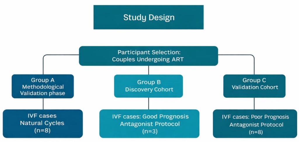
Figure 1. Study design. Participants undergoing assisted reproductive technology (ART) were allocated into three groups: Group A (natural cycle IVF, n=8; methodological validation phase); Group B (good‐prognosis patients undergoing antagonist IVF stimulation protocol, n=3; discovery cohort); and Group C (poor‐prognosis patients, including recurrent implantation failure [RIF], recurrent miscarriage [RM], and advanced maternal age [AMA] cases, undergoing antagonist IVF stimulation protocol, n=8; validation cohort).
Participants were categorized into three groups:
Group A: Methodological validation group (natural cycle patients)
This group included 8 patients undergoing IVF in natural cycles. These patients were included primarily to optimize cumulus cell isolation and miRNA quantification protocols under physiologically controlled conditions.
Group B: Discovery cohort (good prognosis patients)
Three patients undergoing controlled ovarian stimulation were included in this group. Participants were selected based on predefined criteria consistent for good reproductive prognosis. Eligibility criteria included female age ≤35 years, absence of ovulatory disorders, no diagnosis of endometriosis, no evidence of tubal factor infertility, no prior history of recurrent miscarriage or implantation failure, and normal semen parameters according to World Health Organization (WHO) criteria. This cohort was selected to minimize potential biological confounding factors.
Group C: Validation cohort (poor prognosis patients)
Eight patients with poor reproductive prognosis were included in this group. Poor prognosis was defined as the presence of one or more of the following criteria: advanced maternal age (>35 years), history of recurrent implantation failure, or recurrent miscarriage.
This staged design enabled candidate biomarker identification under biologically favorable conditions and validation under clinically heterogeneous conditions. The latter cohort represented clinically relevant populations in which biomarker application would be most relevant. All patients underwent IVF treatment using standardized clinical protocols.
Ovarian stimulation and oocyte retrieval
Patients undergoing controlled ovarian stimulation were treated using GnRH antagonist protocols. Ovarian response was monitored by transvaginal ultrasonography and serum hormone measurements. Final oocyte maturation was induced using human chorionic gonadotropin (hCG).
Oocyte retrieval was performed 34–36 hours following hCG administration using transvaginal ultrasound-guided aspiration. Natural cycle patients underwent oocyte retrieval without ovarian stimulation.
Cumulus cell isolation and Sample processing
Cumulus–oocyte complexes were collected immediately following oocyte retrieval and transferred and incubated for 1 hour in pre-equilibrated culture medium at 37°C under controlled atmospheric conditions (6% CO₂, 5% O₂). Mechanical isolation of cumulus cells was performed under a heated stereomicroscope to ensure preservation of sample integrity and embryo-level traceability. Approximately 50% of the cumulus cell mass was removed mechanically using sterile micro-manipulation tools. Each cumulus cell sample was collected individually into sterile tubes containing RNA stabilization solution (Qiazol Lysis Reagent or RNAprotect, Qiagen) to preserve RNA integrity. Enzymatic removal of residual cumulus cells was subsequently performed using hyaluronidase to allow intracytoplasmic sperm injection (ICSI). Strict traceability was maintained between cumulus cell samples, corresponding oocytes, and resulting embryos. Pooling of samples was deliberately avoided to preserve biological specificity at the individual oocyte level.
RNA extraction and microRNA isolation
Total RNA, including small RNAs, was extracted from individual cumulus cell samples using validated protocols optimized for low-input biological material. Extractions were performed according to the manufacturer’s instructions using commercially available kits specifically designed for small RNA isolation. RNA concentration and quality were assessed prior to downstream analysis. Given the limited cellular material obtained from individual cumulus masses, procedures were carefully optimized to maximize RNA yield and preserve analytical sensitivity. Sample pooling was deliberately avoided to maintain the specific molecular profile of each cumulus–oocyte complex and ensure strict embryo-level traceability.
Apoptosis-related miRNA profiling (Discovery phase)
Apoptosis-related miRNA expression profiling was performed in the discovery cohort using a commercially available human apoptosis-focused miRNA PCR array (Qiagen), enabling simultaneous analysis of 84 apoptosis-associated miRNAs. Total RNA was reverse-transcribed and subjected to pre-amplification prior to quantitative PCR analysis on a real-time PCR platform, according to the manufacturer’s instructions.
The array includes six endogenous controls. Normalization was performed using either the global mean Ct approach or the small nucleolar RNA (SnoRNA) SNORD95, which was identified in our dataset as the most stable reference across samples (Ct variation <1). Relative quantification and data processing were performed using the Qiagen Data Analysis Center. Candidate miRNAs identified in this phase were selected for targeted validation.
Targeted miRNA validation by RT-qPCR (Validation phase)
Expression levels of selected candidate apoptosis-related miRNAs (miR-21, miR-144, miR-146, miR-149) were quantified using reverse transcription quantitative polymerase chain reaction (RT-qPCR). Reverse transcription was performed using TaqMan MicroRNA Reverse Transcription Kit according to manufacturer protocols. Quantitative PCR amplification was performed using TaqMan MicroRNA Assays on a real-time PCR detection system. Each sample was analyzed in accordance with standardized amplification protocols to ensure reproducibility and analytical reliability.
Normalization and relative quantification of miRNA expression
Expression levels of target miRNAs were normalized to U6 small nuclear RNA, which served as an endogenous reference control. Relative miRNA expression was calculated using the ΔCt method: ΔCt = Ct (miRNA) − Ct (U6). Lower ΔCt values correspond to higher relative miRNA expression. Relative expression values were also calculated using the 2^-ΔCt transformation, allowing comparison between samples. This normalization approach is widely used and provides reliable quantification of miRNA expression.
Embryo culture and morphological assessment
Following ICSI, embryos were cultured under standardized laboratory conditions using a time-lapse embryo culture system. Embryo assessment was performed according to established morphological criteria, including cleavage rate, cell number, degree of fragmentation, blastomere symmetry, and overall developmental progression. Based on these parameters, embryos were classified as either good-quality or poor-quality according to standard embryological grading criteria. Each cumulus cell sample was subsequently matched to its corresponding embryo and its developmental outcome.
Statistical analysis
Statistical analysis was performed using Stata version 19 (StataCorp, College Station, TX, USA).
Expression levels were analyzed at multiple analytical levels:
Sample-level analysis: Individual cumulus samples were compared according to embryo quality classification.
Patient-level analysis: Average expression values were calculated per patient and compared between groups.
Paired intra-patient analysis: In patients producing embryos of differing quality, paired comparisons were performed to minimize inter-patient biological variability.
Continuous variables were presented as mean ± standard deviation or as median and interquartile range, as appropriate. Comparisons between independent groups were performed using Student’s t-test. Paired comparisons were performed using paired Student’s t-test.
Statistical significance was defined as p < 0.05, and all tests were two-tailed.
Power analysis: A power analysis was conducted retrospectively to determine the necessary sample size for future large-scale studies. Calculations were based on the mean differences and variance observed in the present pilot data.
Results
Study population and sample characteristics
A total of 19 women undergoing IVF treatment were included in the study. Of these, eight patients underwent natural cycle IVF and constituted the methodological validation group; samples from this cohort were used exclusively to optimize lysis buffer conditions and extraction timing, and to validate assay sensitivity when downscaling from pooled samples to single cumulus cell masses. Three patients were classified as having a good reproductive prognosis and formed the discovery cohort, while eight patients with poor reproductive prognosis comprised the validation cohort. Poor prognosis was defined by the presence of advanced maternal age, recurrent implantation failure, or recurrent miscarriage.
Cumulus cells were successfully isolated from individual oocytes (Table 1) and processed for miRNA analysis. Strict traceability was maintained between each cumulus cell sample and its corresponding oocyte and resulting embryo. All collected samples yielded sufficient RNA for downstream analysis, and miRNA expression was successfully quantified in all analyzed samples, confirming the technical feasibility and analytical reliability of the isolation and quantification protocols.
Technical feasibility and analytical performance
Initial protocol validation was performed using reference miRNAs let-7a and miR-16, selected based on their established stability in cumulus cell literature. The analytical workflow was first optimized using pooled cumulus cells (3 oocytes) and subsequently validated in single-oocyte samples. Following optimization of lysis buffers and incubation times, reproducible detection was achieved in single cumulus masses, confirming the assay’s sensitivity for limited biological input. Subsequently, RT-qPCR amplification was successfully performed for all selected miRNAs (miR-21, miR-144, miR-146 and miR-149), demonstrating the ability to reliably detect and quantify miRNA expression in individual cumulus cell samples. Normalization using U6 endogenous control provided stable reference values across samples, confirming suitability of the normalization strategy. These findings confirm that miRNA expression analysis can be reliably performed on individual cumulus cell samples without pooling, preserving embryo-specific biological information.
Sample-level analysis of miRNA expression according to embryo quality
Comparison of miRNA expression between cumulus cells associated with good-quality and poor-quality embryos was performed at the individual sample level using ΔCt values and relative expression (2^-ΔCt). The results are summarized in Table 2. No statistically significant differences were observed in expression of any of the investigated miRNAs between groups.
However, consistent directional trends were identified:
miR-21 demonstrated lower ΔCt values in cumulus cells associated with poor-quality embryos compared to good-quality embryos, corresponding to higher relative expression. The mean ΔCt difference between poor- and good-quality embryo groups was −0.35 (95% CI: −0.90 to 0.20; p = 0.210). Relative expression (2^-ΔCt) was correspondingly higher in the poor-quality embryo group (mean 1.57 vs 1.18), although this difference did not reach statistical significance (p = 0.123).
miR-146 demonstrated higher ΔCt values in cumulus cells associated with poor-quality embryos compared to good-quality embryos, corresponding to lower relative expression.
The mean ΔCt difference between poor- and good-quality embryo groups was 0.87 (95% CI: −0.41 to 2.15; p = 0.179). Relative expression levels were correspondingly lower in the poor-quality embryo group.
miR-144 expression levels were comparable between groups. Although greater variability was observed in the good-quality embryo group, no statistically significant differences were detected (p = 0.326).
miR-149 expression levels were similar between groups, with minimal differences observed in both ΔCt and relative expression values (p = 0.676) (Table 3).
Patient-level analysis
Patient-level analysis was performed by calculating average expression values per patient. This analysis confirmed findings observed at the sample level, with no statistically significant differences between good- and poor-prognosis patient groups. However, similar directional trends were observed, particularly for miR-21 and miR-146, suggesting potential biological relevance.
Paired intra-patient analysis
Paired analysis was performed in patients producing embryos of differing morphological quality, allowing direct comparison within the same biological environment. This approach reduces confounding effects related to patient-specific biological variability. Paired intra-patient comparisons demonstrated substantial variability in miRNA expression between embryos of differing quality within individual patients. No consistent statistically significant differences were identified. These findings highlight the complex and heterogeneous nature of molecular regulation within individual ovarian follicles.
Biological variability and heterogeneity
Substantial inter-patient and intra-patient variability in miRNA expression was observed across samples. This variability likely reflects biological heterogeneity in follicular development, ovarian physiology, and patient-specific factors. Such variability is consistent with the complex regulatory mechanisms governing folliculogenesis and oocyte competence.
Power analysis and effect size estimation
Power analysis was conducted to estimate sample size requirements necessary to detect observed differences with adequate statistical power. Based on observed effect sizes and variability, substantially larger cohorts would be required to reliably detect statistically significant differences. These findings provide essential information for the design of future adequately powered biomarker validation studies.
Summary of key findings
The key findings of this study indicate that miRNA expression analysis in individual cumulus cell samples is technically feasible and analytically reliable. Although no statistically significant differences were detected between cumulus cells associated with good- and poor-quality embryos in this pilot cohort, consistent directional trends were observed, particularly for miR-21 and miR-146. Notably, substantial biological variability was evident both between patients and within individual patients. Power analysis further suggests that larger cohorts will be required to adequately validate potential associations.
Discussion
The present study was designed as a structured pilot biomarker investigation to evaluate the feasibility, analytical reliability, and preliminary biological relevance of apoptosis-related microRNA expression in human cumulus cells and its association with embryo morphological competence. The findings demonstrate that miRNA profiling can be reliably performed in individual cumulus cell samples obtained during routine IVF procedures and provide important methodological and biological insights relevant to future biomarker development.
A key methodological strength of this study is the successful extraction and quantification of miRNA expression from individual cumulus cell samples without pooling. Given the limited biological material associated with single cumulus–oocyte complexes, preservation of sample individuality ensures biological traceability between cumulus cells, corresponding oocytes, and resulting embryos. Unlike previous studies that relied on pooled samples to increase RNA yield (Karakaya et al., 2012), this approach preserves biological heterogeneity between competent and incompetent embryos and prevents dilution of oocyte-specific molecular signals, consistent with studies emphasizing the importance of individual cumulus–oocyte complex analysis in relation to embryo development [13, 37]. Furthermore, the structured multi-phase design incorporating methodological validation, discovery, and validation cohorts enhances the robustness and translational relevance of the findings, in line with recommended frameworks for staged biomarker evaluation [16]. Inclusion of both good- and poor-prognosis populations provides insight into biomarker behavior across clinically heterogeneous contexts.
No statistically significant differences were identified in the expression of the investigated apoptosis-related miRNAs between cumulus cells associated with good- and poor-quality embryos. However, consistent directional trends were observed, particularly for miR-21 and miR-146. These findings suggest that apoptosis-related miRNAs may reflect subtle differences in follicular and oocyte competence, although the magnitude of these effects appears modest.
The absence of statistically significant differences in the present cohort should be considered within the context of a structured pilot biomarker investigation. The primary objectives of this study were to establish analytical feasibility, characterize biological variability, and generate realistic effect size estimates to inform the design of subsequent validation studies. Biomarker development is inherently multi-phased and requires careful progression from exploratory analyses to adequately powered validation cohorts, as outlined in established biomarker evaluation frameworks [27]. The modest effect sizes observed, together with substantial inter- and intra-patient variability, indicate that considerably larger cohorts will be required both to achieve adequate statistical power and to determine the clinical relevance of the investigated microRNAs.
The use of paired intra-patient analysis represents an additional methodological strength, allowing direct comparison of cumulus cell samples associated with embryos of differing quality within the same patient. This approach minimizes confounding related to inter-patient variability and provides biologically controlled comparisons, as advocated in studies employing paired designs to reduce between-subject noise in high-throughput and molecular investigations [7, 21]. The observed intra-patient heterogeneity further highlights the complexity of follicular biology, consistent with reports demonstrating substantial biological variability that can mask subtle effects in unpaired analyses [25].
Substantial biological variability was observed both between patients and between oocytes from the same patient. Follicular development is a complex and tightly regulated process influenced by genetic, epigenetic, hormonal, and microenvironmental factors, and recent transcriptomic analyses of human cumulus and granulosa cells have demonstrated marked inter- and intra-follicular heterogeneity [23, 41, 42]. This intrinsic heterogeneity likely contributes to the modest group-level differences observed and underscores the challenges inherent in identifying robust molecular biomarkers of oocyte competence.
These findings are broadly consistent with previous studies evaluating cumulus cell molecular profiles in relation to oocyte maturity, fertilization, and embryo development (Table 4). Although associations have been reported, results remain inconsistent across studies, reflecting methodological heterogeneity and limited statistical power. By incorporating structured methodological validation and effect size estimation, the present study provides essential groundwork for future biomarker development.
The selection of apoptosis-related miRNAs was biologically justified by their established roles in ovarian physiology. Apoptosis is central to follicular atresia and regulation of ovarian reserve [28]. miR-21 has been implicated in anti-apoptotic signaling and granulosa cell survival [5] and its increased expression in cumulus cells associated with poor-quality embryos may reflect compensatory responses within compromised follicles. Conversely, miR-146 participates in inflammatory and apoptotic pathway regulation within ovarian tissue [15, 30] and reduced expression may indicate altered follicular homeostasis. Within this biological framework, the observed directional trends appear plausible despite the absence of statistical significance.
This study has several limitations inherent to its pilot design. The sample size was intentionally limited, as the primary objective was methodological validation and effect size estimation rather than definitive biomarker confirmation. Additionally, embryo assessment was based on morphological criteria, which, although widely applied, represent an indirect measure of developmental competence. Future studies incorporating clinical outcomes such as implantation and live birth will be required to fully establish biomarker utility.
Despite these limitations, this study provides essential methodological and biological insight necessary for future biomarker development. The data demonstrate that cumulus cell miRNA profiling is technically feasible, analytically reliable, and capable of capturing biologically meaningful variability. Importantly, realistic effect size estimates and sample size projections are provided to guide adequately powered validation studies.
The development of non-invasive molecular biomarkers capable of predicting embryo competence remains an important goal in reproductive medicine. Cumulus cells represent an attractive source of such biomarkers due to their biological relationship with the oocyte and their accessibility during routine IVF procedures. Future research should focus on larger prospective cohorts, integration of multiple molecular markers, and correlation with clinically relevant outcomes including implantation and live birth. Advances in high-throughput and multi-omics technologies may further enhance biomarker discovery and translational applicability.
Conclusion
This structured pilot study demonstrates that apoptosis-related microRNA expression can be reliably quantified in individual human cumulus cell samples and provides essential methodological validation and biological insight necessary for biomarker development. Although statistically significant associations were not identified in this pilot cohort, consistent directional trends and substantial biological variability were observed. These findings provide critical effect size estimates and establish methodological feasibility, representing essential groundwork for future adequately powered studies aimed at developing non-invasive biomarkers for embryo selection in assisted reproduction.
Funding
This research was funded by Embryogenesis IVF Unit.
Ethics Approval
Greek National Authority of Assisted Reproduction (Protocol No:804).
Competing Interests
The authors declare that they have no competing interests.
References
- Al Edani T, Assou S, Gala A, Ait-Ahmed O, Dechaud H, Hamamah S. Maternal age affects insulin signaling pathway and angiogenic factors of human cumulus cells: importance of MIR-21 and MIR-140. Fertil Steril. 2013; 100(3).
- Almiñana C, Makieva S, Bauersachs S, et al. Endometriosis-associated infertility alters the microRNA signatures of cumulus cells with a particularly pronounced effect in oocytes that failed fertilization. Biol Res. 2025;58(1):62.
- Ambekar AS, Nirujogi RS, Srikanth SM, et al. Proteomic analysis of human follicular fluid: a new perspective towards understanding folliculogenesis. J Proteomics. 2013;87:68-77.
- Andrei D, Nagy RA, van Montfoort A, et al. Differential miRNA Expression Profiles in Cumulus and Mural Granulosa Cells from Human Pre-ovulatory Follicles. MicroRNA. 2018;8(1).
- Assou S, Al-Edani T, Gala A, Haouzi D, Ferrieres A, Hamamah S. Small RNAs: new candidates for the regulation of the human cumulus-oocyte complex crosstalk. Fertil Steril. 2013;100(3).
- Assou S, Haouzi D, Dechaud H, Gala A, Ferrières A, Hamamah S. Comparative gene expression profiling in human cumulus cells according to ovarian gonadotropin treatments. Biomed Res Int. 2013;2013:354582.
- Auer PL, Doerge RW. Statistical design and analysis of RNA sequencing data. Genetics. 2010;185(2):405-416.
- Bartolucci AF, Uliasz T, Peluso JJ. MicroRNA-21 as a regulator of human cumulus cell viability and its potential influence on the developmental potential of the oocyte. Biol Reprod. 2020;103(1).
- Chen D, Zhang Z, Chen B, et al. Altered microRNA and Piwi-interacting RNA profiles in cumulus cells from patients with diminished ovarian reserve. Biol Reprod. 2017;97(1).
- Cui L, Fang L, Mao X, et al. GDNF-Induced Downregulation of miR-145-5p Enhances Human Oocyte Maturation and Cumulus Cell Viability. J Clin Endocrinol Metab. 2018;103(7).
- Da Silva LFI, Da Broi MG, da Luz CM, et al. miR-532-3p: a possible altered miRNA in cumulus cells of infertile women with advanced endometriosis. Reprod Biomed Online. 2021;42(3).
- Feng Y, Qi J, Xue X, et al. Follicular free fatty acid metabolic signatures, and their effects on oocyte competence in non-obese PCOS patients. Reproduction. 2022;164(1):1-8.
- Fragouli E, Wells D, Iager AE, Kayisli UA, Patrizio P. Alteration of gene expression in human cumulus cells as a potential indicator of oocyte aneuploidy. Hum Reprod. 2012;27(8):2559-2568.
- Gardner DK, Schoolcraft WB. In Vitro Culture of Human Blastocyst. In: Jansen R, Mortimer D, editors. Towards Reproductive Certainty. 1999. p.377-388.
- Grieco FA, Sebastiani G, Juan-Mateu J, et al. MicroRNAs regulate expression of proapoptotic proteins. Diabetes. 2017;66(1):100-112.
- Ioannidis JP. Biomarker failures. Clin Chem. 2013;59(1):202-204.
- Karakaya C, Guzeloglu-Kayisli O, Uyar A, et al. Diminished oocyte number in women undergoing IVF is associated with altered microRNA expression in cumulus cells. Fertil Steril. 2012;98(3).
- Karakaya C, Guzeloglu-Kayisli O, Uyar A, et al. Poor ovarian response in women undergoing IVF is associated with altered microRNA expression in cumulus cells. Fertil Steril. 2015;103(6).
- Keller A, Leidinger P, Vogel B, et al. miRNAs can be generally associated with human pathologies as exemplified for miR-144. BMC Med. 2014;12:224.
- Kılıç NÖ, Öner Ç, Kütük D, et al. CCNA2 and CCNB3 as Early Potential Molecular Candidates of Oocyte Maturation in Cumulus-Oophorous Complex Cells from Follicular Fluid. Diagnostics (Basel). 2025;15(20):2658.
- Leek JT, Johnson WE, Parker HS, et al. The sva package for removing batch effects and other unwanted variation in high-throughput experiments. Bioinformatics. 2012;28(6):882-883.
- Liu S, Zhang X, Shi C, et al. Altered microRNAs expression profiling in cumulus cells from patients with PCOS. J Transl Med. 2015;13(1).
- Martínez-Moro Á, González-Brusi L, Lamas-Toranzo I, et al. RNA-sequencing reveals genes linked with oocyte developmental potential in bovine cumulus cells. Mol Reprod Dev. 2022;89(9):399-412.
- McKenzie LJ, Pangas SA, Carson SA, et al. Human cumulus granulosa cell gene expression: a predictor of fertilization and embryo selection in women undergoing IVF. Hum Reprod. 2004;19(12):2869-2874.
- McShane LM, Altman DG, Sauerbrei W, et al. Reporting recommendations for tumor marker prognostic studies (REMARK). J Natl Cancer Inst. 2005;97(16):1180-1184.
- Mendoza C, Ruiz-Requena E, Ortega E, et al. Follicular fluid markers of oocyte developmental potential. Hum Reprod. 2002;17(4):1017-1022.
- Pepe MS, Feng Z, Janes H, et al. Pivotal evaluation of the accuracy of a biomarker used for classification or prediction. J Natl Cancer Inst. 2008;100(20):1432-1438.
- Seifer DB, Cardiner AC, Ferreira KA, Peluso JJ. Apoptosis as a function of ovarian reserve in women undergoing in vitro fertilization. Fertil Steril. 1996;66(4).
- Shafienia H, Montazeri F, Heydari L, et al. Target gene repression mediated by miR-144 and miR-224 in cumulus cells is related to IVF success in PCOS. Reprod Fertil Dev. 2022;34(17): 1089-1098.
- Sirotkin AV, Ovcharenko D, Grossmann R, et al. Identification of microRNAs controlling human ovarian cell steroidogenesis via genome-scale screen. J Cell Physiol. 2009;219(2).
- Su N, Yu X, Duan M, Shi N. Recent advances in methylation modifications of microRNA. Genes Dis. 2025;12(1).
- Tian P, Yan L. Inhibition of microRNA-149-5p induces apoptosis by targeting FASLG. Med Sci Monit. 2016;22.
- Tong XH, Xu B, Zhang YW, et al. Comparative microRNA profiles in human corona radiata cells and cumulus oophorus cells. PLoS One. 2014;9(9).
- Tscherner A, Brown AC, Stalker L, et al. STAT3 signaling stimulates miR-21 expression in bovine cumulus cells during in vitro oocyte maturation. Sci Rep. 2018;8(1):11527.
- Velthut-Meikas A, Simm J, Tuuri T, et al. Small RNA-seq of human granulosa cells reveals miRNAs in FSHR and aromatase genes. Mol Endocrinol. 2013;27(7).
- Vigo FM, Fraietta R, Rodrigues F, et al. Cumulus cell microRNA expression when LH is added to ovarian stimulation protocol: a pilot study. Reprod Biomed Online. 2021;43(6).
- Wathlet S, Adriaenssens T, Segers I, et al. Cumulus cell gene expression predicts better cleavage-stage embryo or blastocyst development and pregnancy. Hum Reprod. 2011;26(5):1035-1051.
- Woo I, Christenson LK, Gunewardena S, et al. Micro-RNAs involved in cellular proliferation have altered expression profiles in granulosa cells of young women with diminished ovarian reserve. J Assist Reprod Genet. 2018;35(10).
- Wright EC, Hale BJ, Yang CX, et al. MicroRNA-21 and PDCD4 expression during in vitro oocyte maturation in pigs. Reprod Biol Endocrinol. 2016;14:21.
- Wu H, Zhu R, Zheng B, et al. Single-Cell Sequencing Reveals an Intrinsic Heterogeneity of the Preovulatory Follicular Microenvironment. Biomolecules. 2022;12(2):231.
- Wu YT, Tang L, Cai J, et al. High bone morphogenetic protein-15 level in follicular fluid is associated with high quality oocyte and embryonic development. Hum Reprod. 2007; 22(6):1526-1531.
- Wyse BA, Fuchs Weizman N, Kadish S, et al. Transcriptomics of cumulus cells – a window into oocyte maturation in humans. J Ovarian Res. 2020;13(1):93.
- Xu B, Zhang YW, Tong XH, Liu YS. Characterization of microRNA profile in human cumulus granulosa cells. Mol Cell Endocrinol. 2015;404: 26-36.
- Yang H, DeWan AT, Desai MM, Vermund SH. Preimplantation genetic testing for aneuploidy: challenges in clinical practice. Hum Genomics. 2022;16(1):69.
- Yerushalmi GM, Salmon-Divon M, Ophir L, et al. Characterization of the miRNA regulators of the human ovulatory cascade. Sci Rep. 2018;8(1).