Original Article
HJOG 2026, 25 (4), 376-388 | doi: 10.33574/hjog.0644
Eleni Nazou1,†, Despoina Mavrogianni2,†, Anastasios Potiris1, Efthalia Moustakli3, Ekaterini Domali4, Periklis Panagopoulos1, George Daskalakis4, Peter Drakakis1, Sofoklis Stavros1
1Third Department of Obstetrics and Gynecology, University General Hospital “ATTIKON”, Medical School, National and Kapodistrian University of Athens, Athens, Greece
2First Department of Obstetrics and Gynecology, Alexandra Hospital, Medical School, National and Kapodistrian University of Athens, Athens, Greece
3Department of Nursing, School of Health Sciences, University of Ioannina, 4th Kilometer National Highway Str. Ioannina-Athens, Ioannina, Greece
4First Department of Obstetrics and Gynecology, Alexandra Hospital, Medical School, National and Kapodistrian University of Athens, Athens, Greece
†These authors contributed equally to this work.
Correspondence: Eleni Nazou, Third Department of Obstetrics and Gynecology, University General Hospital “ATTIKON”, Medical School, National and Kapodistrian University of Athens, 124 62 Athens, Greece. E-mail: nazoueleni@gmail.com

Abstract
Follicular fluid (FF) and semen contain microRNAs (miRNAs), important regulators of reproductive physiology and potential non-invasive biomarkers of infertility. This study examined miR-99a and miR-143 expression in infertile couples undergoing assisted reproductive technology (ART) and their correlations with reproductive characteristics in sperm and FF. Paired FF and semen samples were collected from 36 couples. Quantitative real-time PCR was used to quantify normalized miRNA expression, with G6PD as the internal reference gene. Non-parametric statistical tests were applied to assess correlations between clinical, hormonal, and embryological data, as well as to perform comparative analyses between sexes and tissue types. Significant differences in miR-99a and miR-143 expression were observed between sperm and follicular fluid samples, with higher expression in male than female samples (p < 0.001 and p = 0.004, respectively). MiRNA expression was significantly associated with selected female reproductive parameters, particularly AMH and FSH levels, whereas no correlations were identified with LH, sperm motility or total sperm count. Moreover, a significant correlation was observed between miR-143 expression and embryo quality, with higher levels in embryos of superior morphology (p = 0.046). These findings indicate differential expression of miR-99a and miR-143 between sperm and follicular fluid and suggest their involvement in early embryonic development and gametogenesis. Specifically, in ART, miR-143 may represent a potential marker of embryo quality, although this finding should be interpreted cautiously due to the limited sample size. Further studies are needed to validate these findings and clarify the underlying mechanisms.
Keywords: miR-99a, miR-143, follicular fluid, sperm, microRNAs, embryo quality, assisted reproduction.
Introduction
A major global health problem, infertility affects 8–12% of couples who are of reproductive age globally. Male factors contribute to approximately 50% of infertility cases overall and account for 20–30% as the primary cause, underscoring the need to assess both partners in clinical practice [1,2]. A significant percentage of infertility cases remain unexplained despite advancements in assisted reproductive technologies (ART), underscoring the need for better genetic indicators and a deeper comprehension of reproductive biology [3].
Small non-coding RNAs, microRNAs (miRNAs) control post-transcriptional gene expression and are essential for various biological functions, including reproduction [4,5]. In the ovary, follicular fluid miRNAs contribute to folliculogenesis, steroidogenesis, oocyte competence and intercellular communication [6,7].
Notably, miR-99a and miR-143 are important regulators of ovarian function. MiR-99a targets the insulin-like growth factor 1 receptor (IGF-1R) to influence granulosa cell proliferation and death, leading to aberrant follicular formation [8]. Similar to this, miR-143 is crucial for granulosa cell physiology because it inhibits the generation of estradiol generated by follicle-stimulating hormone (FSH) and targets KRAS, a key element of the MAPK signaling pathway [9,10]. These findings demonstrate the role of miRNAs in several signaling pathways crucial for ovarian homeostasis, including PI3K/AKT and MAPK.
In the male reproductive system, miRNAs are critical regulators of spermatogenesis and sperm function. Altered miRNA expression can disrupt key molecular pathways, affecting meiosis, chromatin remodeling, and germ cell differentiation [11,12]. Furthermore, members of the miR-99 family exhibit altered expression in the testes of men with non-obstructive azoospermia, suggesting their involvement in male infertility [14,15]. In addition, miR-143 has been associated with sperm maturation and function, further supporting the role of miRNAs as regulators of male reproductive capacity [16].
This study evaluated miR-99a and miR-143 expression in paired sperm and follicular fluid samples from infertile couples and examined their associations with semen quality, hormonal profiles and embryo development, while accounting for tissue- specific and sex related variation.
Materials and Methods
Study Population
All samples were obtained from the Assisted Reproduction Unit of “Attikon” University Hospital (Athens, Greece). The study protocol was approved by the Institutional Ethics Committee, and written informed consent was obtained from all participants in accordance with the Declaration of Helsinki.
A total of 36 infertile couples (36 women and 36 men) undergoing ART (IVF/ICSI) between September 2024 and January 2025 were included. Female inclusion criteria were: age 25–45 years, regular menstrual cycles (21–35 days), normal hormonal profile according to WHO guidelines, and the presence of both ovaries. All participants had a history of at least two years of infertility and at least one previous unsuccessful IVF attempt.
Women underwent a short GnRH agonist protocol for controlled ovarian stimulation (COS). No participant had received hormonal treatment for at least three months prior to study inclusion. Male participants provided semen samples following 3–5 days of sexual abstinence. All participants were screened for infectious diseases prior to sample collection. Baseline demographic and clinical characteristics, including age, body mass index (BMI), and duration of infertility, were recorded before initiation of ovarian stimulation. Female and male characteristics are presented in Tables 1 and 2, respectively.
Ovarian stimulation was performed using individualized gonadotropin (Gn) protocols based on ultrasound monitoring and hormonal assessment. Dosage and duration were adjusted until the day of human chorionic gonadotropin (hCG) administration.
On the day of oocyte retrieval, FF samples were collected following transvaginal aspiration. Purified samples were either used immediately or stored at −80 °C until further analysis.
Semen samples were collected by masturbation into sterile containers and allowed to liquefy for 30 minutes at 37 °C. Analysis was performed according to WHO guidelines. Samples were washed with PBS and stored at −80 °C until RNA extraction (within one week).
RNA Isolation, Reverse Transcription, and Quantitative Real-Time PCR
Total RNA was extracted using the Nucleic Acid Extraction Rapid Kit (magnetic bead-based method; Jiangsu Bioperfectus Technologies Co., Ltd.) according to the manufacturer’s instructions. RNA samples were stored at −80 °C until further processing.
Complementary DNA (cDNA) synthesis was performed using the LunaScript RT SuperMix Kit (New England BioLabs, #E3010L). The resulting cDNA samples were stored at −20 °C.
Quantitative real-time PCR (qPCR) was conducted using Luna Universal qPCR Master Mix (New England BioLabs, #M3003) on a LightCycler 480 Real-Time PCR System (Roche Applied Science). The G6PD gene was selected as an internal control based on its stable expression across the analyzed sample types. Primer sequences are presented in Table 4. Normalized expression values were used for comparative analyses. All RNA extraction and amplification procedures were performed under standardized experimental conditions according to the manufacturers’ instructions. G6PD was selected as the reference gene based on its stable amplification profile and consistent Ct values across the analyzed sample types.
Statistical Analysis
Statistical analyses were performed using SPSS software (version 20.0; IBM Corp., Armonk, NY, USA). Comparisons between two independent groups were conducted using the non-parametric Mann–Whitney U test. Associations between semen parameters and miRNA expression were evaluated using Spearman’s rank correlation coefficient, whereas correlations involving female reproductive parameters were assessed using Spearman’s rank correlation coefficient due to the non-parametric distribution of the data and the limited sample size. Adjusted correlation analyses were performed to control for the effects of age, body mass index (BMI), smoking status, and number of retrieved oocytes. Comparisons among more than two groups were performed using the non-parametric Kruskal–Wallis test, followed by multiple pairwise comparisons using Dunn’s procedure with Bonferroni correction. A p-value < 0.05 was considered statistically significant.
Study Population and Sample Collection
A total of 36 infertile couples were included in the study. FF samples were collected from women during oocyte retrieval, and semen samples were obtained from their male partners. Expression levels of miR-99a and miR-143 were analyzed in all samples. The distribution of embryo quality categories is presented in Table 3.
Differential Expression of miR-99a and miR-143 in Sperm and Follicular Fluid Samples
Significant differences in the expression levels of miR-99a and miR-143 were observed between sperm and follicular fluid samples (Figure 1, Table 5). Specifically, miR-99a expression differed significantly between sperm (6.64 ± 2.46) and FF samples (2.37 ± 1.51; p < 0.001), while miR-143 also showed significantly different expression levels between sperm and FF samples (4.57 ± 2.02 vs 3.43 ± 2.15; p = 0.004).
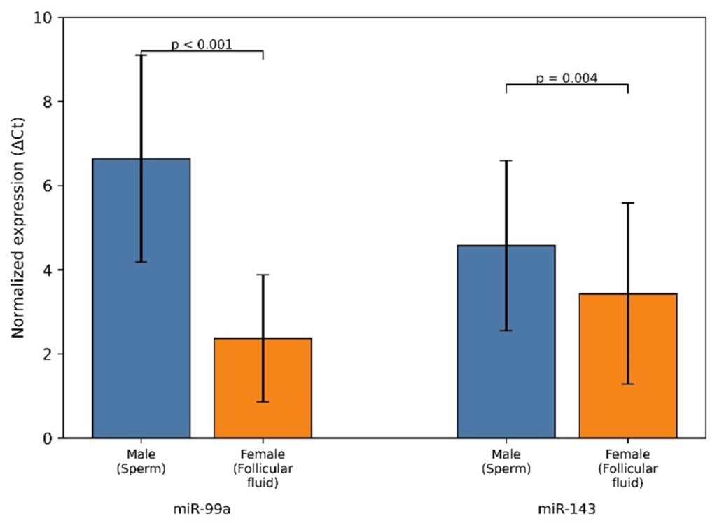
Figure 1. Comparison of miR‐99a and miR‐143 expression levels. Bar charts represent normalized miRNA expression values in male (sperm) and female (FF) samples. Significant differences were observed between sperm and follicular fluid samples for both miR‐99a and miR‐143.
Association between miRNA Expression and Semen Parameters
No statistically significant correlations were identified between miR-99a expression and sperm motility (rs = -0.146, p = 0.395) or total sperm count (rs = 0.166, p = 0.334). Similarly, miR-143 expression showed no significant associations with semen parameters (p > 0.05) (Table 6).
Subgroup analysis revealed that men with lower sperm motility (≤42%) exhibited higher mean miR-99a and miR-143 levels compared to those with higher motility (>42%); however, these differences were not statistically significant (p = 0.203 and p = 0.214, respectively). Likewise, men with higher total sperm counts (>39 million) showed slightly increased miRNA expression levels, without statistical significance (p = 0.222 and p = 0.168). The motility group comparisons are presented in Table 6B.
Association between miRNA Expression and Female Clinical Parameters
In the female group, significant positive correlations were observed between miR-99a or miR-143 expression levels and AMH concentrations. In addition, miR-143 expression was positively associated with FSH levels and negatively correlated with the number of retrieved oocytes. No significant associations were found with age, body mass index (BMI), duration of infertility, LH, estradiol or prolactin (p > 0.05). Results for miR-99a and miR-143 are presented in Tables 7 and 8, respectively. Adjusted correlations between hormonal parameters and miR-99a and miR-143 are presented in Table 9 and Table 10.
Association between miRNA Expression and Embryo Quality
No statistically significant differences were observed in miR-99a expression across embryo quality groups (Poor, Average, Good, Excellent; p = 0.436). Embryo quality was assessed on Day 3 according to standard morphological criteria based on blastomere symmetry, fragmentation rate, and cell number. In contrast, miR-143 expression differed significantly among these groups (p = 0.046).
Post hoc Bonferroni analysis demonstrated that embryos of excellent quality exhibited significantly higher miR-143 expression levels compared with those of poor quality (p = 0.042) (Table 11).
Summary of Statistical Findings
No statistically significant differences were observed in miR-99a expression across embryo quality groups (Poor, Average, Good, Excellent; p = 0.436). In contrast, miR-143 expression differed significantly among these groups (p = 0.046). A summary of statistical associations is presented in Table 12.
Discussion
This study demonstrated differential expression of miR-99a and miR-143 between sperm and follicular fluid samples from ART patients, likely reflecting tissue-specific and sex-related differences. Limited associations were observed with female hormonal parameters(AMH and FSH), while higher miR-143 expression was positively associated with embryo quality.
The observed differences in miR-99a and miR-143 expression between sperm and follicular fluid samples are consistent with previous studies highlighting the role of miRNAs in spermatogenesis [5,12]. Dysregulated miRNAs, which regulate key processes such as meiosis, germ cell development, and chromatin remodeling, may contribute to male infertility [17,18]. The genes targeted by these microRNAs, including FGFR3, SMAD3, AKT3, and TGFBR1, are associated with signaling pathways that regulate germ cell survival and differentiation[19]. These pathways are essential for maintaining spermatogonial stem cell self-renewal and preventing apoptosis during early germ cell development, thereby ensuring proper progression through spermatogenesis. Moreover, miR-143 is enriched in epididymis tissues, while miR-99a is present in seminal plasma exosomes, supporting their role in intercellular communication [16].
Despite these molecular links, no correlation was found between the expression of these miRNAs and standard semen analysis parameters, including sperm motility and total number of spermatozoa. This apparent contradiction suggests that these miRNAs may exert more pronounced effects at the molecular level or through processes not detected by conventional semen analysis. In fact, previous studies have shown that sperm function can be impaired through signaling pathways such as calcineurin signaling without necessarily altering classical semen parameters [20,21].
Both miR-99a and miR-143 are also involved in female reproductive processes, including steroidogenesis, granulosa cell function, and folliculogenesis [4,6]. While miR-143 regulates granulosa cell proliferation and estrogen secretion through inhibition of components in the MAPK pathway, miR-99a regulates folliculogenesis and ovarian signaling pathways. In particular, miR-99a has been shown to target IGF-1R, a key regulator of granulosa cell survival and proliferation, thereby influencing follicular development and oocyte maturation. Additionally, studies indicate that miR-143-3p induces granulosa cell apoptosis and contributes to follicular dysfunction in PCOS [22,23].
The functional relevance of miR-143 in male reproduction may also be supported by its evolutionary conservation across species, suggesting a fundamental regulatory role. Previous studies have demonstrated that miR-143-3p interacts with key regulators of spermatogenesis, including CREBBP, a transcriptional coactivator involved in chromatin remodeling and energy metabolism during early germ cell development [24]. In this context, circular RNA circ-CREBBP has been shown to act as a molecular sponge for miR-143-3p, thereby modulating the expression of genes associated with cell survival and sperm function, such as MCL1 and CREB1. Given that MCL1 is an anti-apoptotic member of the BCL-2 family, these interactions suggest a role for miR-143 in regulating sperm survival and motility. Indeed, dysregulation of circ-CREBBP and its associated pathways has been linked to impaired sperm motility and increased apoptosis, further supporting the involvement of miR-143 in male reproductive function [25].
In the present study, significant correlations were observed between miRNA expression and selected female reproductive parameters, particularly AMH levels. In contrast, no significant associations were identified with age, BMI, LH, estradiol, prolactin, or duration of infertility. Despite this, a correlation was observed between miR-143 expression and embryo morphology, with higher levels in embryos displaying better morphology. Our results are consistent with previous reports indicating that FF miRNAs influence oocyte developmental potential through intracellular pathways and granulosa cell activity [7,26–29]. These miRNAs are capable of modulating gene expression within granulosa cells after uptake, thereby altering the follicular microenvironment and influencing oocyte competence. Thus, miR-143 appears to be associated with early embryonic development through modulation of the follicular microenvironment. However, these findings should be interpreted cautiously, as the small subgroup sizes, particularly in the poor and excellent embryo quality groups, limit the robustness of subgroup comparisons.
Interestingly, significant positive correlations were observed between both miR-99a and miR-143 expression levels and AMH concentrations. These associations remained statistically significant even after adjustment for potential confounding factors, including age, BMI, smoking status, and number of retrieved oocytes. Since AMH reflects ovarian reserve and granulosa cell function, these findings may support the involvement of both miRNAs in follicular physiology and ovarian microenvironment regulation. MiR-99a and miR-143 regulate granulosa cell function via IGF-1R, MAPK, and TGF-β-related pathways.
In addition, miR-143 expression demonstrated a positive association with FSH levels and a negative correlation with the number of retrieved oocytes. Although the underlying mechanisms remain unclear, these findings may indicate a relationship between miR-143 expression and ovarian response during ART cycles. Higher miR-143 levels in women with lower oocyte yield could reflect altered follicular microenvironment dynamics or compensatory regulatory mechanisms associated with diminished ovarian responsiveness.
No statistically significant correlations were observed between miRNA expression and age, BMI, LH, estradiol, prolactin, or duration of infertility. This may suggest that the observed miRNA alterations are closely associated with specific ovarian functional pathways rather than with general demographic or endocrine characteristics.
All results support the view that miRNAs act as active regulators in the reproductive system rather than merely as diagnostic markers of physiological conditions [30]. Through major cellular pathways such as PI3K/AKT, TGF-β/SMAD, and MAPK/ERK, miRNAs may influence various functions in reproductive physiology. Such physiological processes may include follicular development, spermatogenesis, and even embryo implantation. The process of fertilization and early embryogenesis might be negatively affected if the complex interactions are somehow impaired.
Although miR-143 showed an association with embryo quality, further validation in larger cohorts is required before any potential clinical application can be considered. The complexity of miRNA regulation is evident from its lack of association with clinical indicators. This indicates that the usefulness of miRNAs as biomarkers is contingent upon their combined use with other biomarkers [3].
This study is limited by its small size, lack of causal inference, evaluation of only two miRNAs, absence of lifestyle confounder assessment and independent validation, and limited adjustment for clinical confounders.
Further research should focus on large-scale, multicenter studies integrating clinical, transcriptomic, and proteomic data. Advanced approaches such as next-generation sequencing (NGS) may provide deeper insights into reproductive miRNA networks. Further studies should investigate the mechanisms governing miRNA transport, stability, and intercellular communication in the reproductive system. Advances in miRNA biology may facilitate the development of targeted diagnostic and therapeutic approaches for infertility.
Conclusions
In conclusion, miR-99a and miR-143 expression differed significantly between sperm and follicular fluid samples, with miR-143 showing positive associations with embryo quality and morphology, and selected female hormonal and ovarian response parameters, but not with conventional semen parameters.
These findings suggest that miR-99a and miR-143 may regulate gametogenesis and early embryonic development, with miR-143 representing a potential biomarker of embryo quality pending further validation.
Furthermore, RT-PCR-based methods are used in the majority of recent investigations, including this one. NGS may make it possible to find more pertinent miRNAs.
These results require more confirmation. Larger, more diverse populations, combined with molecular and clinical research, will be necessary to clarify the mechanisms by which miRNAs influence human fertility and reproductive outcomes.
Author Contributions
Conceptualization, E.N. and S.S.; methodology, D.M., E.M. and E.D.,validation, D.M., P.C. and A.P.; investigation, E.M., and E.N.; writing—original draft preparation, E.N. and S.S.; writing—review and editing, D.M., A.P., E.M., P.P., E.D., G.D. and P.D.; visualization, S.S.; supervision, P.D.; project administration, P.D. All authors have read and agreed to the published version of the manuscript.
Funding
This research received no external funding.
Institutional Review Board Statement
The study was conducted in accordance with the Declaration of Helsinki and approved by the Institutional Ethics Committee.
Informed Consent Statement
Written informed consent was obtained from all subjects involved in the study.
Data Availability Statement
The data presented in this study are available on reasonable request from the corresponding author. Due to ethical and privacy considerations related to patient data, the datasets are not publicly available.
Acknowledgments
The authors would like to acknowledge the contribution of the clinical and laboratory staff involved in the assisted reproduction procedures.
Conflicts of Interest
The authors declare no conflicts of interest.
References
- Vander Borght M, Wyns C. Fertility and infertility: definition and epidemiology. Clin Biochem. 2018; 62:2-10. doi:10.1016/j.clinbiochem.2018.03.012.
- Nachtigall RD. International disparities in access to infertility services. Fertil Steril. 2006;85:871-5. doi:10.1016/j.fertnstert.2005.08.066.
- Carson SA, Kallen AN. Diagnosis and management of infertility: a review. JAMA. 2021;326:65. doi:10.1001/jama.2021.4788.
- Maalouf SW, Liu WS, Pate JL. MicroRNA in ovarian function. Cell Tissue Res. 2016;363:7-18. doi:10.1007/s00441-015-2307-4.
- Hossain MM, Sohel MMH, Schellander K, Tesfaye D. Characterization and importance of microRNAs in mammalian gonadal functions. Cell Tissue Res. 2012;349:679-90. doi:10.1007/s00441-012-1469-6.
- Montazerian M, Yasari F, Aghaalikhani N. Ovarian extracellular microRNAs as potential non-invasive biomarkers: an update. Biomed Pharmacother. 2018; 106:1633-40. doi:10.1016/j.biopha.2018.07.073.
- Sohel MMH, Hoelker M, Noferesti SS, et al. Exosomal and non-exosomal transport of extracellular microRNAs in follicular fluid: implications for bovine oocyte developmental competence. PLoS One. 2013; 8:e78505. doi:10.1371/journal.pone.0078505.
- Geng Y, Sui C, Xun Y, Lai Q, Jin L. miRNA-99a can regulate proliferation and apoptosis of human granulosa cells via targeting IGF-1R in polycystic ovary syndrome. J Assist Reprod Genet. 2019; 36:211-21. doi:10.1007/s10815-018-1335-x.
- Zhang L, Zhang X, Zhang X, Lu Y, Li L, Cui S. miRNA-143 mediates the proliferative signaling pathway of FSH and regulates estradiol production. J Endocrinol. 2017; 234:1-14. doi:10.1530/JOE-16-0488.
- Du X, Zhang L, Li X, Pan Z, Liu H, Li Q. TGF-β signaling controls FSHR signaling-reduced ovarian granulosa cell apoptosis through the SMAD4/ miR-143 axis. Cell Death Dis. 2016;7:e2476. doi: 10.1038/cddis.2016.379.
- Walker WH. Regulation of mammalian spermatogenesis by miRNAs. Semin Cell Dev Biol. 2022; 121:24-31. doi:10.1016/j.semcdb.2021.05.009.
- Salas-Huetos A, James ER, Aston KI, Carrell DT, Jenkins TG, Yeste M. The role of miRNAs in male human reproduction: a systematic review. Andrology. 2020;8:7-26. doi:10.1111/andr.12714.
- Abu-Halima M, Hammadeh M, Backes C, et al. Panel of five microRNAs as potential biomarkers for the diagnosis and assessment of male infertility. Fertil Steril. 2014;102:989-97.e1. doi: 10.1016/j.fertnstert.2014.07.001.
- Yao C, Yuan Q, Niu M, et al. Distinct expression profiles and novel targets of microRNAs in human spermatogonia, pachytene spermatocytes, and round spermatids between OA patients and NOA patients. Mol Ther Nucleic Acids. 2017; 9:182-94. doi:10.1016/j.omtn.2017.09.007.
- Vojtech L, Woo S, Hughes S, et al. Exosomes in human semen carry a distinctive repertoire of small non-coding RNAs with potential regulatory functions. Nucleic Acids Res. 2014; 42:7290-304. doi:10.1093/nar/gku347.
- Guo J, Grow EJ, Yi C, et al. Chromatin and single-cell RNA-seq profiling reveal dynamic signaling and metabolic transitions during human spermatogonial stem cell development. Cell Stem Cell. 2017; 21:533-46.e6. doi:10.1016/j.stem.2017.09.003.
- Von Kopylow K, Staege H, Spiess AN, et al. Differential marker protein expression specifies rarefaction zone-containing human Adark spermatogonia. Reproduction. 2012;143:45-57. doi: 10.1530/REP-11-0290.
- Chitiashvili T, Hsu F, Dror I, Plath K, Clark A. FGFR3 is expressed by human primordial germ cells and is repressed after meiotic initiation to form primordial oocytes. Stem Cell Reports. 2022; 17:1268-78. doi:10.1016/j.stemcr.2022.04.015.
- Miyata H, Satouh Y, Mashiko D, et al. Sperm calcineurin inhibition prevents mouse fertility with implications for male contraception. Science. 2015; 350:442-5. doi:10.1126/science.aad0836.
- Bray C, Son JH, Kumar P, Meizel S. Mice deficient in CHRNA7, a subunit of the nicotinic acetylcholine receptor, produce sperm with impaired motility. Biol Reprod. 2005; 73:807-14. doi: 10.1095/biolreprod.105.042184.
- Cao J, Huo P, Cui K, et al. Follicular fluid-derived exosomal miR-143-3p/miR-155-5p regulate follicular dysplasia by modulating glycolysis in granulosa cells in polycystic ovary syndrome. Cell Commun Signal. 2022;20:61. doi: 10.1186/s12964-022-00876-6.
- Zhao Y, Pan S, Li Y, Wu X. Exosomal miR-143-3p derived from follicular fluid promotes granulosa cell apoptosis by targeting BMPR1A in polycystic ovary syndrome. Sci Rep. 2022;12:4359. doi:10.1038/s41598-022-08423-6.
- Boussouar F, Goudarzi A, Buchou T, et al. A specific CBP/p300-dependent gene expression programme drives the metabolic remodelling in late stages of spermatogenesis. Andrology. 2014; 2:351-9. doi:10.1111/j.2047-2927.2014.00184.x.
- Ding N, Zhang Y, Huang M, et al. Circ-CREBBP inhibits sperm apoptosis via the PI3K-Akt signaling pathway by sponging miR-10384 and miR-143-3p. Commun Biol. 2022;5:1339. doi: 10.1038/s42003-022-04263-2.
- Noferesti SS, Sohel MMH, Hoelker M, et al. Controlled ovarian hyperstimulation induced changes in the expression of circulatory miRNA in bovine follicular fluid and blood plasma. J Ovarian Res. 2015;8:81. doi:10.1186/s13048-015-0208-5.
- Gebremedhn S, Gad A, Ishak GM, et al. Dynamics of extracellular vesicle-coupled microRNAs in equine follicular fluid associated with follicle selection and ovulation. Mol Hum Reprod. 2023; 29:gaad009. doi:10.1093/molehr/gaad009.
- Xiao Y, Peng X, Peng Y, et al. Macrophage-derived extracellular vesicles regulate follicular activation and improve ovarian function in old mice by modulating local environment. Clin Transl Med. 2022;12:e1071. doi:10.1002/ctm2.1071.
- Tesfaye D, Worku D, Rings F, et al. Identification and expression profiling of microRNAs during bovine oocyte maturation using heterologous approach. Mol Reprod Dev. 2009;76:665-77. doi:10.1002/mrd.21005.
- Dai M, Hong L, Yin T, Liu S. Disturbed follicular microenvironment in polycystic ovary syndrome: relationship to oocyte quality and infertility. Endocrinology. 2024;165:bqae023. doi: 10.1210/endocr/bqae023.
- Nadri P, Nadri T, Gholami D, et al. Role of miRNAs in assisted reproductive technology. Gene. 2024; 927:148703. doi:10.1016/j.gene.2024.148703.